

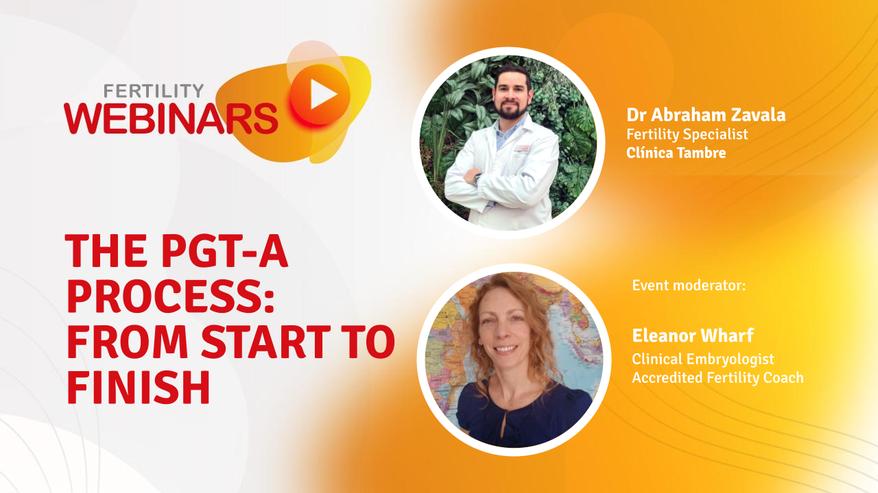
Curious about how PGT-A really works — from the biopsy to the final embryo result? In this live event, Dr Abraham Zavala, Fertility Specialist at Clínica Tambre Alicante, breaks down the entire PGT-A process step by step.
The session was hosted by Eleanor Wharf, Clinical Embryologist & Accredited Fertility Coach, recently awarded Best Fertility Coach 2025 by the European Fertility Society.
In this webinar, Dr Abraham Zavala explained the full preimplantation genetic testing process, from the biological rationale behind PGT-A to how embryos are biopsied, analysed, and ultimately selected for transfer.
PGT-A, or preimplantation genetic testing for aneuploidy, is designed to assess whether an embryo has the correct number of chromosomes. As Dr Zavala explained, this information can help reduce the risk of miscarriage and improve the chance of success per embryo transfer, particularly in specific patient groups.
He emphasised from the outset that PGT-A is a diagnostic tool, not a treatment. “There is no test in fertility that can guarantee a successful pregnancy,” he explained, but PGT-A can provide additional information to guide decision-making.
Preimplantation genetic testing (PGT) is a group of tests used to analyse the genetic makeup of embryos created through IVF. While many people use the term PGT to mean PGT-A, Dr Zavala clarified that there are three distinct types of PGT, each with a different purpose.
PGT-A focuses on chromosome number. PGT-M is used to detect a specific single-gene condition. PGT-SR looks at structural rearrangements of chromosomes, such as translocations.
A genetically normal embryo, he explained, is not the same as a good-quality embryo under the microscope. “A beautiful embryo that is genetically abnormal has a lower chance of implanting than a lower-quality embryo with a normal genetic result.”
PGT-A is the most commonly used form of genetic testing in IVF. It checks whether an embryo has 46 chromosomes. Embryos with more or fewer chromosomes are classified as aneuploid.
PGT-M is used when one or both parents carry a known genetic mutation that can cause a serious inherited disease. In this case, the test looks for that specific gene.
PGT-SR is used when a parent has a structural chromosomal rearrangement, such as a balanced translocation. Although the parent may be healthy, embryos can inherit an unbalanced chromosome arrangement, which can lead to implantation failure or miscarriage.
Each test has a different indication, and Dr Zavala stressed that PGT should never be used routinely without a clear reason.
Dr Zavala outlined the main clinical situations where PGT-A is commonly considered. These include women over the age of 37, couples with recurrent pregnancy loss, repeated implantation failure, or known chromosomal abnormalities in one of the parents.
He explained that aneuploidy increases with age, largely due to changes in egg quality. Under the age of 35, around 60–70% of embryos may be chromosomally normal. Over the age of 40, that number drops significantly, with only around two out of ten embryos expected to be euploid.
PGT-A does not increase the overall chance of pregnancy across multiple transfers, but it can increase the chance of success per transfer and reduce the likelihood of miscarriage caused by chromosomal abnormalities.
PGT-A is an invasive procedure, and Dr Zavala spent time explaining how it is performed and why timing matters.
Today, embryos are biopsied at the blastocyst stage, usually on day 5 or 6 of development. At this stage, the embryo has two distinct cell lines: the inner cell mass, which becomes the fetus, and the trophectoderm, which becomes the placenta.
Only cells from the trophectoderm are biopsied.
We never take cells from the inner cell mass.
Dr Zavala explained. This approach reduces the risk to the embryo and improves the reliability of results.
Once the biopsy is taken, embryos must be frozen while genetic analysis is performed. For this reason, all PGT-A cycles involve frozen embryo transfer.
Historically, genetic testing was sometimes performed on day-three embryos. Dr Zavala explained why this approach is no longer recommended.
At day three, embryos have very few cells. Removing one or two cells represents a significant proportion of the embryo and may compromise development. In addition, all cells at this stage are still undifferentiated, so it is unclear which cells would have contributed to the fetus or placenta.
Blastocyst-stage biopsy is now considered safer and more informative.
Most laboratories today use next-generation sequencing (NGS) to analyse embryo biopsies. NGS allows rapid and detailed assessment of chromosome number.
PGT-A results generally fall into three categories: euploid, aneuploid, or mosaic.
A euploid embryo has 46 chromosomes. An aneuploid embryo has too many or too few chromosomes. Mosaic embryos contain two different cell lines, one normal and one abnormal.
Dr Zavala explained that even euploid embryos can contain a very small proportion of abnormal cells. Mosaic embryos are defined by a higher percentage of abnormal cells, typically between 15 and 50%, depending on laboratory thresholds.
Mosaic embryos are one of the most complex and debated areas of PGT-A. Dr Zavala explained that mosaic embryos can result in healthy pregnancies, but they have lower implantation rates and slightly higher miscarriage rates compared to euploid embryos.
In cases where mosaic embryos are transferred, closer prenatal monitoring is recommended. This may include invasive testing such as amniocentesis, rather than relying only on non-invasive screening.
In Spain, mosaic embryos are usually transferred only when no euploid embryos are available and after detailed counselling.
Dr Zavala outlined how PGT is regulated in Spain. Spanish law permits PGT in assisted reproduction, but the level of regulation depends on the type of test.
PGT-A is permitted and largely guided by clinical recommendation and patient choice, provided appropriate counselling is given. PGT-M is more tightly regulated and is generally approved only for serious, early-onset genetic diseases.
Some conditions are pre-approved, while others require individual authorisation from the national committee for assisted reproduction. This approval process can take six to twelve months and may involve genetic testing of extended family members.
Dr Zavala was clear that PGT-A is not a perfect test. It is invasive and, like all medical tests, has false positives and false negatives.
A small percentage of embryos classified as aneuploid may actually be capable of developing into healthy pregnancies. For this reason, the risks and benefits must always be weighed carefully.
The main benefit of PGT-A is improved embryo selection, particularly for women of advanced maternal age. It can reduce miscarriage risk and shorten the time to pregnancy by avoiding transfers of embryos with little chance of success.
Recent evidence shows mixed results for routine PGT-A use. In women under 35, studies have not shown a clear benefit. In women over 37, outcomes appear more favourable, although data must be interpreted carefully.
Dr Zavala highlighted that many studies are biased, as only women who produce blastocysts can undergo PGT-A. This can exaggerate success rates when compared to untested cycles.
Professional societies currently recommend PGT-A as an optional add-on rather than routine treatment, with careful patient selection.
To illustrate real-world use, Dr Zavala shared several clinical cases involving PGT-M, PGT-SR, and PGT-A.
These included couples with inherited genetic disease, chromosomal translocations, and women of advanced age. In each case, PGT helped identify embryos with the best chance of leading to a healthy pregnancy, though not every transfer resulted in success.
These examples reinforced a key message: PGT provides information, not guarantees.
Dr Zavala described a typical case involving a woman of advanced maternal age undergoing IVF. As he explained, the likelihood of chromosomal abnormalities increases significantly with age, largely due to changes in egg quality.
In this case, several embryos were created and cultured to the blastocyst stage. PGT-A testing revealed that only one embryo was euploid, while the others were aneuploid or mosaic. Without PGT-A, embryo selection would have relied solely on morphological appearance, which does not reliably predict chromosomal normality.
By identifying the euploid embryo, the clinic was able to prioritise that embryo for transfer, reducing the risk of miscarriage associated with aneuploid embryos. However, Dr Zavala emphasised that even a euploid embryo does not guarantee implantation or live birth, as other factors such as uterine receptivity and maternal health still play a role.
This case illustrated how PGT-A can be useful in shortening the time to pregnancy by avoiding transfers of embryos with very low implantation potential, particularly in women over 37.
Another case involved a couple with a history of recurrent pregnancy loss. Standard investigations had not identified a clear anatomical or hormonal cause, and previous embryos had not been genetically tested.
In this situation, PGT-A was introduced to assess whether embryonic chromosomal abnormalities might be contributing to the miscarriages. Several embryos reached the blastocyst stage, and genetic testing showed that most were aneuploid, with only one euploid embryo available for transfer.
Dr Zavala explained that while PGT-A cannot prevent miscarriage from all causes, it can help reduce miscarriage risk when chromosomal abnormalities are a contributing factor. In this case, transferring a euploid embryo allowed the clinical team to focus attention on uterine and maternal factors if implantation failed, rather than repeatedly transferring embryos with a high likelihood of genetic abnormality.
This case demonstrated how PGT-A can be a diagnostic tool that helps clarify the underlying causes of repeated pregnancy loss, even if it does not guarantee success.
Dr Zavala also discussed a case involving a woman aged 39 with endometriosis who had undergone IVF with PGT-A. One euploid embryo and one low-level mosaic embryo were identified.
The euploid embryo was transferred first, but unfortunately resulted in a second-trimester pregnancy loss. This raised the question of whether the remaining mosaic embryo could still be considered for transfer.
Dr Zavala explained that mosaic embryos generally have lower implantation rates and a slightly higher risk of miscarriage than euploid embryos. However, it is uncommon for low-level mosaic embryos to result in chromosomally abnormal fetuses. For this reason, in the absence of euploid embryos, transferring a low-level mosaic embryo may be considered after thorough counselling.
In such cases, invasive prenatal testing, such as amniocentesis, is recommended if pregnancy is achieved. This case highlighted both the complexity of mosaic results and the importance of individualised decision-making when no euploid embryos remain.
Dr Zavala presented an example involving a couple in which one partner carried a balanced chromosomal translocation. Although the carrier was healthy, this type of rearrangement increases the risk of producing embryos with unbalanced chromosomes, leading to implantation failure or miscarriage.
In this case, PGT-SR was used to identify embryos with a balanced or normal chromosomal structure. Importantly, Dr Zavala noted that PGT-A is typically performed alongside PGT-SR to ensure that embryos are not only free of the specific rearrangement but also chromosomally normal overall.
Several embryos were excluded due to unbalanced chromosomal results. The remaining embryos were prioritised for transfer based on both genetic findings and morphological quality. This case illustrated how PGT-SR can prevent repeated unsuccessful transfers and reduce miscarriage risk in couples with known chromosomal rearrangements.
Finally, Dr Zavala described a case involving a known single-gene disorder. One or both partners were carriers of a genetic condition that could result in a serious inherited disease.
In this situation, PGT-M was used to identify embryos that did not carry the disease-causing mutation. As Dr Zavala explained, PGT-M requires extensive preparation, including genetic testing of the couple and, in some cases, other family members. This process can take several months and requires approval from regulatory authorities in Spain.
In this case, embryos that were free of the mutation were identified and considered suitable for transfer. PGT-A was also performed to exclude embryos with major chromosomal abnormalities. This example highlighted the role of PGT-M in preventing transmission of serious genetic disease, while also demonstrating the complexity and time commitment involved.
Across all examples, Dr Zavala consistently emphasised that PGT provides information rather than certainty. Genetic testing can help reduce risk, guide embryo selection, and clarify causes of failure, but it cannot guarantee implantation, pregnancy, or live birth.
These cases also reinforced the importance of:
careful patient selection for PGT
detailed genetic counselling
realistic expectations
individualised treatment planning
As Dr Zavala explained, the value of PGT lies not in replacing clinical judgement, but in supporting it with additional, carefully interpreted information.
A mosaic embryo is classified as either low-level or simple mosaic. Low-level mosaicism refers to a situation where there is a low percentage of an additional abnormal cell line. The exact threshold depends on laboratory reference values, but it is usually below 30–40%.
A simple mosaic refers to how many chromosomes are affected. When we analyse an embryo, we are only analysing a small group of cells, not the entire embryo. Because of this, retesting can theoretically result in a different outcome.
However, retesting is not something we routinely recommend. Retesting requires thawing the embryo, biopsying it again, refreezing it, and then thawing it once more for transfer. This process may negatively affect implantation potential.
In the absence of euploid embryos, a low-level mosaic embryo is one that we would consider transferring after careful counselling. Some embryos may be suitable for re-biopsy, depending on their quality and the number of cells available, but this must always be evaluated on an individual basis.
There is evidence suggesting that embryos which undergo a second biopsy tend to have lower implantation rates. However, it is not entirely clear why.
It may be related to repeated freezing and thawing, or to the loss of additional cells during the second biopsy. We cannot determine the exact cause, but this potential reduction in implantation potential is one of the reasons why retesting is approached cautiously.
Accuracy depends partly on the genetic testing company used. Different clinics work with different laboratories, and academic research methods are often more precise than commercial testing.
Based on published evidence, embryos that are classified as aneuploid but are actually euploid occur in approximately 2% of embryos. With commercial testing, this percentage may be slightly higher.
Mosaicism contributes to this uncertainty, as results are not always fully reproducible.
I am very sorry to hear about the pregnancy loss. Based on clinical experience, mosaic embryos have a lower chance of implantation, a lower chance of developing into a healthy pregnancy, and a slightly higher risk of miscarriage compared to euploid embryos.
That said, it is uncommon for a low-level or simple mosaic embryo to result in a chromosomally abnormal fetus. There is a theoretical risk, which is why we recommend invasive prenatal testing, such as amniocentesis, if pregnancy is achieved.
In Spain, mosaic embryos are typically transferred only when no euploid embryos are available. Patients may choose to undergo another IVF cycle before transferring a mosaic embryo. This decision should be discussed carefully with the treating clinic.
Yes. At our clinic, we provide genetic counselling when discussing abnormal karyotypes, mosaic embryos, or genetic matching results.
In addition, most genetic testing laboratories also offer genetic counselling services upon request. This is an important step in helping patients understand the potential implications of specific genetic findings.
When laboratories perform PGT-M or PGT-SR, they usually perform PGT-A at the same time. This is because we want to avoid transferring embryos that are free of a specific genetic condition but have a major chromosomal abnormality.
PGT-M and PGT-SR often involve additional testing of family members, which increases the cost. This step is not required for PGT-A.
In most cases, only one embryo biopsy is needed, and all required testing can be done from that single sample. Thawing embryos solely for additional testing is generally avoided if possible.
The significance of a deletion depends on its location, the genes involved, and the percentage of the affected cell line. This information should be discussed with a genetic counsellor.
It is also important to remember that in mosaic embryos, the deletion is present only in the secondary cell line. The proportion of that cell line is a key factor in risk assessment.
In general, PGT-A is not recommended for donor-egg embryos because it does not appear to improve pregnancy rates.
However, in cases of recurrent miscarriage or repeated implantation failure, PGT-A may be considered to help exclude embryonic chromosomal causes and focus attention on uterine or maternal factors.
In practice, this can be difficult, as clinics often outsource genetic testing. Patients can ask which laboratory is used and request information about misdiagnosis rates and quality assurance measures.
These laboratories should be able to provide data on accuracy and error rates. Clinics should also be transparent about which companies they work with.
Embryo biopsy is a highly specialised and delicate procedure. Not every embryologist is trained to perform it.
The procedure involves using a laser to create a small opening in the embryo and removing cells from the trophectoderm at the correct stage of development. Timing and technical skill are critical.
In many clinics, only senior embryologists perform biopsies. In some cases, genetic testing companies send experienced embryologists to clinics to ensure biopsies are carried out by trained professionals.
Like ICSI, embryo biopsy requires extensive training, practice, and ongoing quality control to minimise harm to the embryo.


+ 3 more answers

+ 1 more answers

Necessary cookies are absolutely essential for the website to function properly. This category only includes cookies that ensures basic functionalities and security features of the website. These cookies do not store any personal information.
Analytical cookies are used to understand how visitors interact with the website. These cookies help provide information on metrics the number of visitors, bounce rate, traffic source, etc.
| Cookie | Duration | Description |
|---|---|---|
| _ga | 2 years | This cookie is installed by Google Analytics. The cookie is used to calculate visitor, session, campaign data and keep track of site usage for the site's analytics report. The cookies store information anonymously and assign a randomly generated number to identify unique visitors. |
| _gat_UA-38575237-21 | 1 minute | No description |
| _gid | 1 day | This cookie is installed by Google Analytics. The cookie is used to store information of how visitors use a website and helps in creating an analytics report of how the website is doing. The data collected including the number visitors, the source where they have come from, and the pages visted in an anonymous form. |
Any cookies that may not be particularly necessary for the website to function and is used specifically to collect user personal data via analytics, ads, other embedded contents are termed as non-necessary cookies. It is mandatory to procure user consent prior to running these cookies on your website.
Other uncategorized cookies are those that are being analyzed and have not been classified into a category as yet.
| Cookie | Duration | Description |
|---|---|---|
| _gat_FSQM52 | 1 minute | No description |
| cf_ob_info | No description | |
| cf_use_ob | No description |